
Page 239 - Vibrational Spectroscopic Imaging for Biomedical Applications
P. 239
Raman Detection of Car otenoids in Human T issue 215
In Fig. 7.13, we summarize the main results of a comparison of MP
distributions and concentrations obtained with Raman imaging and
24
nonmydriatic lipofuscin fluorescence excitation, respectively.
Figure 7.13a and b compare images of both methods, obtained for
the same subject. Compared to the RRI image, the fluorescence-
based image is nearly identical, with the exception of a smoother
appearance of the distribution. This is due to the derivation of the
MP density map as the logarithm of a ratio between perifoveal and
foveal fluorescence intensities, which tends to slightly compress the
“dynamic range” of the density map amplitudes, and smoothen out
the resulting MP distribution. For a subgroup of 16 subjects, we
integrated the MP levels of images obtained with both methods for
each individual over the whole macula region, and plot the results
in Fig. 7.13c. Using a best fit that is not forced through zero, we
obtain a high-correlation coefficient of R = 0.89 between both meth-
ods. Forcing the fit through zero, the correlation coefficient drops
slightly to R = 0.80. The high correlation is remarkable in view of the
completely different optical beam paths and derivation methods
used to calculate MP densities in both methods.
7.6 Raman Detection of Carotenoids in
Living Human Skin
Levels of carotenoids are much lower in the skin relative to the mac-
ula of the human eye, but higher light excitation intensities and lon-
ger acquisition times can be used in Raman detection approaches to
compensate for this drawback. Since the bulk of the skin carotenoids
are in the superficial layers of the dermis, and since the concentra-
tions are relatively low, the thin-film Raman equation given above
should still be a good approximation.
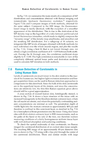
A cross section of excised human skin, histologically stained, is
shown in Fig. 7.14. It shows a layer structure of the tissue with an
increased homogeneity in the bloodless stratum corneum layer, where
the cell nuclei are absent, and where the potentially confounding mel-
anin concentrations are minimal as well. The penetration depth of
visible light into the stratum corneum is approximately 400 μm and
therefore is confined to this outermost layer, as sketched in Fig. 7.14
for a hemispherical beam penetration into the tissue. Using skin tissue
sites with thick stratum corneum layer in RRS measurements, such as
the palm of the hand or the sole of the foot, one therefore realizes
measuring conditions of a fairly homogeneous uniform tissue layer
with well-defined absorption and scattering conditions.
To check the spatial uniformity of skin carotenoids, we Raman
imaged the carotenoid distribution of an excised skin tissue sample.
The resulting RRI image of an approximately 40-μm diameter skin
area is shown in Fig. 7.15 along with a line plot through the center of
the carotenoid distribution. These and similar results with large