
Page 56 - Vibrational Spectroscopic Imaging for Biomedical Applications
P. 56
Algal Cells, Cartilage, and IRENI 33
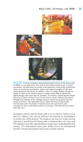
FIGURE 2.2 A series of photos showing illuminated mirrors in the beam path
for IRENI. In the right photo, the array of illuminated toroids (nine of twelve
are shown), the fi rst array of mirrors in the beamline, refocus the synchrotron
beam as it exits the synchrotron. Notice two bright rectangles of light just
above and below the center. In addition, there is a large shadow across the
center of each mirror, which is due to a water-cooled tube to absorb the
higher energy soft x rays and UV radiation. The bottom, central picture shows
the array of plane mirrors (11 of 12 are shown), which redirects the beams
through exit windows of the ultrahigh vacuum system housing the two fi rst
arrays of mirrors. The upper-left picture shows the fi nal 24 illuminated
mirrors that collect the swath of radiation from the synchrotron and
recombine it into a collimated bundle of 12 beams. (Printed with permission
from Ref. 21.)
final plane mirrors and the beam path in air are shown. Notice on the
first two mirrors, one can see shadows surrounded by illumination
on either side of the shadow. The shadows are due to a water-cooling
tube that is installed in the beam path to eliminate the soft x rays and
UV radiation from the extracted beam to prevent overheating the first
mirrors. The illuminated beam path clearly shows the recombined
bundle of beams leaving the final series of plane mirrors.