
Page 61 - Vibrational Spectroscopic Imaging for Biomedical Applications
P. 61
1000 60
1500
2000 40
2500 cm –1 section @ 3500 cm –1 μm
3000 20
3500
0.8 0.6 0.4 0.2 0.0 –0.2 0
60 40 μm 20 0
Absorbance
1000 60
1500
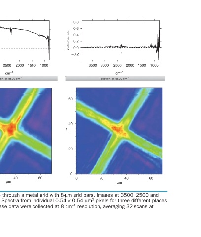
2000 40 Color-scale images of transmission at the IRENI beamline through a metal grid with 8-μm grid bars. Images at 3500, 2500 and
2500 cm –1 section @ 3500 cm –1 μm 1500 cm –1 are shown, where red/blue indicates high/low absorption. Spectra from individual 0.54 × 0.54 μm 2 pixels for three different places
3000 20 in the field of view are also shown to demonstrate the noise level. These data were collected at 8 cm –1 resolution, averaging 32 scans at
3500
1.0 0.5 0.0 Absorbance –0.5 60 40 μm 20 0 0
1000 60
1500
2000 40
2500 cm –1 section @ 3500 cm –1 μm
3000
20
3500 2.5 kHz data collection rate.
FIGURE 2.5
1.5 1.0 0.5 0.0 0
0
60 40 μm 20
Absorbance
37