
Page 64 - Vibrational Spectroscopic Imaging for Biomedical Applications
P. 64
40 Cha pte r T w o
Wavelength (μm)
2.5 3 4 5 6 8 10
2.4
Diamond
2.3
Index of Refraction 2.2 BaF 2
ZnS
1.5
1.4
1.3 CaF 2
4000 3000 2000 1000
–1
Wavenumber (cm )
FIGURE 2.8 Indices of refraction in the mid-IR region for common window
31
31
32
materials including diamond, ZnS, BaF , and CaF 32 that illustrate the
2 2
relative low dispersion of diamond. (Printed with permission from Ref. 18.)
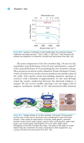
The main components of the flow chamber (Fig. 2.9) are two dia-
mond films (typical thickness 0.4 to 0.8 μm), hold apart by a spacer 34
with a typical thickness of 15 μm defining the flow chamber volume.
They are grown on silicon wafers (diameter 32 mm, thickness 0.5 mm),
which are etched away locally at seven positions around the center of
the wafer. This exposes seven free-standing diamond openings or
windows with a diameter of approximately 2.5 mm each through
which the sample, sandwiched between the diamond windows, is
imaged. We use several smaller openings over one bigger hole to
improve mechanical stability of the sub-micrometer-thin diamond
Lid
Base
Top Seal
Water Layer Silicon
with Sample Wafer
Diamond Spacer
Films (2×) Diamond Silicon
Film
Seals (2×) Silicon Spacer Water Tubes (2×) Wafer
Water Tubes (2×) Wafers (2×) Bottom Seal
Base
FIGURE 2.9 Design details of the fl ow chamber. Left panel: Three-quarter
section view. Center panel: Explosion view indicating the water fl ow (blue
arrows). Color codes for both panels: lid (blue), base (green), diamond
(yellow) coated silicon wafers (gray), spacer (red), seals (purple), water tube
(cyan), and screws (gray) fastening the lid to the base. Right panel:
Photograph of the fl ow chamber (top view). The ends of the water in/outlet
tubes have luer connectors for easy connecting to other equipment, e.g.,
a pump. (Printed with permission from Ref. 18.)