
Page 62 - Vibrational Spectroscopic Imaging for Biomedical Applications
P. 62
38 Cha pte r T w o
60
100
80
40
60
μm
40
20
20
0
0
0 20 40 60
μm (b)
(a)
FIGURE 2.6 Unprocessed IR (a) and visible (b) grey-scale images of an
agglomeration of 6- μm polystyrene beads. The IR image is generated from
–1
the absorption at 3025 cm and 100 on the grey scale that corresponds to
0.125 absorption units. This was measured in transmission at IRENI.
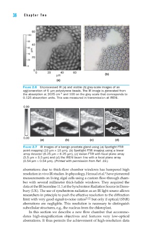
0.90
0.00
(a) (b) (c) (d)
FIGURE 2.7 IR images of a benign prostate gland using (a) Spotlight FTIR
point mapping (10 μm × 10 μm), (b) Spotlight FTIR imaging using a linear
array detector (6.25 μm × 6.25 μm), (c) Varian FTIR with focal plane array
(5.5 μm × 5.5 μm) and (d) the IRENI beam line with a focal plane array
(0.54 μm × 0.54 μm). (Printed with permission from Ref. 19.)
aberrations due to thick-flow chamber windows has hampered high
6
resolution in vivo IR studies. In phycology, Heraud et al. have pioneered
measurements on living algal cells using a custom flow-through cham-
ber with several millimeter thick-halide windows. They acquired the
data at the IR beamline 11.1 at the Synchrotron Radiation Source in Dares-
bury (UK). The use of synchrotron radiation as an IR light source allows
researchers in principle to push the effective resolution to the diffraction
limit with very good signal-to-noise ratios 21,22 but only if optical/(SNR)
aberrations are negligible. This resolution is necessary to distinguish
subcellular structures, e.g., the nucleus from the chloroplast.
In this section we describe a new flow chamber that accommo-
dates high-magnification objectives and features very low-optical
aberrations. It thus permits the achievement of high-resolution data