
Page 102 - Advances in Biomechanics and Tissue Regeneration
P. 102
98 6. REVIEW OF THE ESSENTIAL ROLES OF SMCS IN ATAA BIOMECHANICS
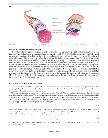
Adventitia
External elastic lamina
Media
Internal elastic lamina
Intima
FIG. 6.2 Structure of the arterial wall. Courtesy of T.C. Gasser, Structure and Basic Properties of the Arterial Wall, 2017 (Indisponible en Accès Libre).
6.2.1.2 A Multilayered Wall Structure
The aortic wall is divided into three main layers surrounding the lumen where the blood flow circulates (Fig. 6.2).
Each layer has its function and proper mechanical properties [6, 14, 27, 57, 61, 62]. The adventitia, which is the most
external layer, contains fibroblasts and is particularly collagen-rich, according to its protective role for the entire wall
against high stress. The internal layer, called the intima, is directly in contact with the blood flow. It also constitutes a
selective barrier of endothelial cells for preventing the wall from blood product infiltration and delivering oxygen and
nutrients from the blood to the internal wall. The inner medial layer is separated from adventitia and intima by two
elastic laminae, and represents about two-thirds of the whole thickness of the wall. All these layers have a passive
mechanical response to the loading induced by the blood flow, but only the media can also act actively due to the
presence of contractile SMCs. The media is structured into several MLUs (Fig. 6.1)[21, 62], where a layer of SMCs
is tight between two thin elastin sheets through a complex network of interlamellar elastin connections [57]. The SMCs
are oriented in the direction of the ECM fibers in order to better transmit the forces to each other and to successive
MLUs. The number of MLUs varies according to the diameter of the artery [62] and the size of the organism: 6 8
for mice and 40 70 for the human body [21].
6.2.2 Basics of Aortic Biomechanics
It is commonly assumed that only the adventitia and the media are involved in the mechanical response of the entire
wall, neglecting the mechanical role of the intima. This assumption is not valid in the case of pathologies resulting in a
thickening of the intima such as atherosclerosis.
The aorta is submitted to four types of mechanical stresses (Fig. 6.3). The two main components are the axial one, σ z ,
and the circumferential one, σ θ . The two other components are, namely σ r (radial stress) and τ w (wall shear stress). The
wall shear stress results from the friction of the blood onto the wall. The circumferential stress is related to the disten-
sion of the aorta with the variation of the blood pressure. It can reach about 150 kPa under normal conditions [21].
It can be approximated by the Laplace law according to:
P r
(6.1)
σ θ ¼
t
where P is the blood pressure, r the internal aortic radius, and t the thickness of the wall. If the number of MLUs varies
according to the arterial diameter and across species [63], the average tension per MLU was shown to remain constant
at T ¼ 2 N/m [21], and its average circumferential stress can be determined by
T
(6.2)
σ θ ¼
t MLU
As the mean thickness of an MLU is about t MLU ’ 15μm, it was estimated that the average normal circumferential stress
across the aorta is σ θ ¼ 133 kPa [21].
I. BIOMECHANICS