
Page 104 - Advances in Biomechanics and Tissue Regeneration
P. 104
100 6. REVIEW OF THE ESSENTIAL ROLES OF SMCS IN ATAA BIOMECHANICS
350.0
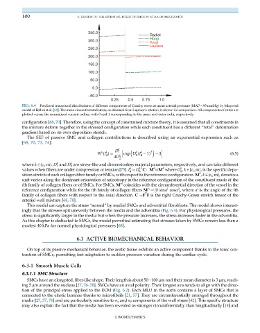
Radial
Hoop
300.0
Axial
Laplace
250.0
200.0
150.0
100.0
50.0
0.0
–50.0
0.25 0.5 0.75 1.0
FIG. 6.4 Predicted transmural distributions of different components of Cauchy stress at mean arterial pressure (MAP 93 mmHg) by bilayered
model of Bellini et al. [68]. The mean circumferential stress, as obtained from Laplace’s relation, is shown for comparison. All components of stress are
plotted versus the normalized current radius, with 0 and 1 corresponding to the inner and outer radii, respectively.
configuration [68, 70]. Therefore, using the concept of constrained mixture theory, it is assumed that all constituents in
the mixture deform together in the stressed configuration while each constituent has a different “total” deformation
gradient based on its own deposition stretch.
The SEF of passive SMC and collagen contributions is described using an exponential expression such as
[68, 70, 73, 74]:
D k h i
k k 1 k k 2 1 (6.5)
4 2 4
4D
W ðI Þ¼ k exp D ðI 1Þ
2
k
k
where k 2{c i , m}. D and D are stress-like and dimensionless material parameters, respectively, and can take different
1 2 2
k
k
k
k
k
values when fibers are under compression or tension [75]. I ¼ G C : M
M where G , k 2{c i , m}, is the specific depo-
4 h h
k
sition stretch of each collagen fiber family or SMCs, with respect to the reference configuration. M , k 2{c i , m}, denotes a
unit vector along the dominant orientation of anisotropy in the reference configuration of the constituent made of the
m
ith family of collagen fibers or of SMCs. For SMCs, M coincides with the circumferential direction of the vessel in the
i
i
i
reference configuration while for the ith family of collagen fibers M ¼½0 sinα cosα , where α is the angle of the ith
c i
T
family of collagen fibers with respect to the axial direction. C ¼F F is the right Cauchy-Green stretch tensor of the
arterial wall mixture [68, 70].
This model can capture the stress “sensed” by medial SMCs and adventitial fibroblasts. The model shows interest-
ingly that the stresses spit unevenly between the media and the adventitia (Fig. 6.4). For physiological pressures, the
stress is significantly larger in the media but when the pressure increases, the stress increases faster in the adventitia.
As this chapter is dedicated to SMCs, the model permitted estimating that stresses taken by SMCs remain less than a
modest 40 kPa for normal physiological pressures [68].
6.3 ACTIVE BIOMECHANICAL BEHAVIOR
On top of its passive mechanical behavior, the aortic tissue exhibits an active component thanks to the tonic con-
traction of SMCs, permitting fast adaptation to sudden pressure variation during the cardiac cycle.
6.3.1 Smooth Muscle Cells
6.3.1.1 SMC Structure
SMCs have an elongated, fiber-like shape. Their length is about 50 100 μm and their mean diameter is 3 μm, reach-
ing 5 μm around the nucleus [27, 76–78]. SMCs have an axial polarity. Their longest axis tends to align with the direc-
tion of the principal stress applied to the ECM (Fig. 6.2). Each MLU in the aorta contains a layer of SMCs that is
connected to the elastic laminae thanks to microfibrils [21, 57]. They are circumferentially arranged throughout the
media [27, 57, 76] and are particularly sensitive to σ z and σ θ components of the wall stress [42]. This specific structure
may also explain the fact that the media has been revealed as stronger circumferentially than longitudinally [14] and
I. BIOMECHANICS