
Page 106 - Advances in Biomechanics and Tissue Regeneration
P. 106
102 6. REVIEW OF THE ESSENTIAL ROLES OF SMCS IN ATAA BIOMECHANICS
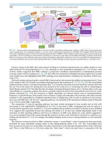
FIG. 6.6 Summary of the main signaling pathways involved in SMC contractility and phenotypic switching. α-SMA, alpha smooth muscle actin;
Ang II, angiotensin II; AT1, angiotensin receptor; C v , caveola; PDGF, platelet-derived growth factor; PDGFRβ, PDGF receptor; ROCK, Rho kinase
involved in cytoskeleton turnover; TGF-β, beta transforming growth factor; TGFBR1/2, TGF-β receptor. Specific genes mainly involved in the loss of
contractility—ACTA2: encoding the α-SMA, MYH11: encoding the myosin heavy chains. Shape and orientation of an SMC according to its ECM. The
SMCs can synchronize their contraction along their strongest axis thanks to intercellular interactions: between several SMCs through gap junctions,
or between endothelial cells from the intima and innermost MLUs of SMCs through vasoactive agonists, neurotransmitters, or secreted GAGs.
Calcium entries in the SMC after some stimuli resulting in membrane depolarization are widely studied in vitro
with electrical [95], electrochemical [39, 50, 100], chemical, or even mechanical stimulation [47, 101]. In fact, some
of these studies suggested that SMCs undergo a progressive membrane depolarization as intraluminal pressure
increases under normal conditions [47, 48, 101]. But when the mechanical stimulation becomes higher than normal,
some studies have also highlighted that SMCs undergo more depolarization, resulting in an alteration of their reac-
tivity [102].
+
The most common protocol used to control SMC contraction in vitro remains the addition of potassium ions K from
a KCl solution with a 50–80 mM concentration that depolarizes the membrane [39, 49, 50, 100, 102]. The extracellular
2+
media must also contain a calcium concentration [Ca ] e to cause the activation of myosin heads by calcium entry into
the cell. This is the reason for adding the CaCl 2 solution to the media [49], or immersing the cells in a physiological
Krebs-Ringer solution [39]. The latter has the advantage of keeping biological tissues alive. Calcium entry is also reg-
ulated thanks to a cytosolic oscillator that allows a periodic release of calcium from intracellular reservoirs (i.e., endo-
plasmic reticulum) [46, 103]. The frequency is highly dependent on external stimuli such as neurotransmitters,
hormones, or growth factors. If its primary role is to induce a single cell contraction, the secondary role of the cytosolic
oscillator is also responsible for membrane depolarization of neighboring cells in order to synchronize the contrac-
tion of several SMCs [46, 55]. The Ca 2+ signaling pathway was included in the mechanical cellular model of Murtada
et al. [104] to model SMC contractility.
The angiotensin II (Ang II) signaling pathway has been widely developed in mice models and its link with
aneurysms is well explained by Malekzadeh et al. [105]. It may lead to SMC contraction and may be used as a
vasoconstrictor agonist in mice models [15, 105] or for isolated cells by addition in a bath [52]. But the review of Michel
et al. suggests that angiotensin II may also damage the intima [15]. In this case, intimal degradation leads to the
activation of other signaling pathways that have an influence on SMC tone. Moreover, some studies suggested a
very active biological role of the intima through the secretion of nitric oxide (NO) that is involved in a pathway con-
trolling cell relaxation [52, 102]. Accordingly, the intimal integrity seems to have a strong influence on cell contractile
response.
The filament overlap involved in SMC contraction creates a “cross-bridge” whose function has been previously
described by some subcellular models based on the sliding-filament theory [106, 107]. The cross-bridges have
been assumed to be based on contractility activation/deactivation cycles through phosphorylation of the contractile
unit. That is what Dillon et al. [108] have called the “latch state,” which was used later in association with the
I. BIOMECHANICS