
Page 105 - Advances in Biomechanics and Tissue Regeneration
P. 105
6.3 ACTIVE BIOMECHANICAL BEHAVIOR 101
that the forces produced by the SMCs are maximized in this direction [76]. This ability of endothelial cells and SMCs to
align along the direction of the applied stress has been confirmed by a number of in vitro studies [79–81].
The arrangement of SMCs in the media used to be controversial [82]. The most recent studies (1980s [63, 83], 1990s
[84], and 2000 [85]) describe SMC orientation as circumferential whereas a helical and oblique disposition was reported
earlier (1960s [86], 1970s [87]). Fujiwara and Uehara showed an oblique orientation in 1992 [76]. Likewise, data are
controversial about alignment parallel to the vessel surface: Clark and Glagov [83] agree with this statement, unlike
Fujiwara and Uehara [76]. Furthermore, some authors mention a change of SMC orientation in each subsequent MLU,
creating a herringbone-like layout [85, 88]. Humphrey suggested that the SMCs are oriented helically, closer to a cir-
cumferential direction [27], but O’Connell suggested the SMCs may also be slightly radially tilted [82].
A recent study pointed out the importance of the helical disposition, suggesting that SMCs are oriented according to
two intermingled helices [89]. This disposition was assumed in several tissue models [90, 91]. Moreover, a tissue model
for coronaries taking into account the orientation of SMCs suggested they contribute both to circumferential and axial
stresses and tend to reorient toward the circumferential direction when blood pressure is increased [92]. Other studies
[83, 93] suggested that the almost circumferential orientation is only valid for inner MLUs of the ascending thoracic
aorta because SMCs seem to orient more axially close to the adventitia. This pattern was also confirmed by Fujiwara
and Uehara [76].
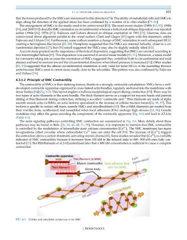
6.3.1.2 Principle of SMC Contractility
The contractility of SMCs is their defining feature, thanks to a strongly contractile cytoskeleton. SMCs have a well-
developed contractile apparatus organized in cross-linked actin bundles, regularly anchored into the membrane with
dense bodies [94](Fig. 6.5). This layout implies a bulbous morphological aspect during contraction [95]. There may be
two types of actin filaments in the same bundle. The thick filament serves as a support for myosin heads and permits
sliding of thin filaments during contraction, defining a so-called “contractile unit.” Thin filaments are made of alpha
smooth muscle actin (α-SMA), an actin isoform specialized in the increase of cellular traction forces [24, 96, 97]. This
isoform is specific to certain cell types, namely SMCs and myofibroblasts [45]. The α-SMA filaments are created from
their rod-like form, synthesized, and assembled when focal adhesions (FAs) undergo high stresses [24, 96]. Genetic
mutations may affect the genes encoding the components of the contractile apparatus (Fig. 6.6) and lead to ATAAs
(Table 6.1).
The main signaling pathways controlling SMC contraction are summarized in Fig. 6.6. More details about these
pathways may be found in Refs. [26, 34, 46, 48, 51, 98]. However, it is important to mention that SMC contractility
2+
is controlled by the modulation of intracellular ionic calcium concentration [Ca ] i . The SMC membrane has many
2+
invaginations called caveolae where extracellular Ca 2+ ions can enter the cell [99]. The increase of [Ca ] i triggers
2+
the contraction above a certain threshold, activating myosin chains [48]. Some studies revealed that [Ca ] i is a reliable
indicator of SMC contractility because it increases from 100 nM in the relaxed state to 600 800 nM once fully con-
tracted [27]. But Hill-Eubanks et al. [48] underlined later that a 400 nM concentration is sufficient to cause a complete
contraction.
FIG. 6.5 Cellular and subcellular architecture of the SMC.
I. BIOMECHANICS