
Page 111 - Advances in Biomechanics and Tissue Regeneration
P. 111
6.4 MECHANOSENSING AND MECHANOTRANSDUCTION 107
Because SMCs are dynamic systems, their cytoskeleton remains in constant evolution during cellular processes. It
consists in a dense fibrous actin structure that allows the cell for shape maintenance and generation of traction forces
required notably during migration (Fig. 6.8). The cytoskeleton of SMCs is particularly rich in contractile α-SMA thin
filaments (see Section 6.3.1) that are used to enhance the traction forces required for cell function. SMC contraction
involves a quick remodeling of its cytoskeleton in order to recruit contractile thin filaments in the direction of applied
forces [24, 96] and to follow its change of shape while renewing noncontractile cortical structures [52].
In summary, the SMC may be considered a powerful sensor of the mechanical state across the aortic wall. The high
sensitivity of SMCs led many research teams to point out their implication in arterial disease, including aortic aneu-
rysms [20, 22, 27, 36, 42, 77, 129].
6.4.2 The Key Role of SMCs in ATAAs
The role of SMCs in the development of ATAAs is now well accepted [57, 76].
Several studies have already mentioned the change of SMC behavior in cardiovascular disease, and the conse-
quences on the arterial wall. It was shown that hypertension is perceived by SMCs as permanent stimuli through
the increase of wall stress, which induces collagen synthesis to reinforce the wall resulting in an increasing thickness
[15, 27, 98, 131].
In atherosclerosis and restenosis, the growth of plaques between the media and the intima is due to SMC prolifer-
ation and migration toward the intima, forming a neointima [40, 129]. The neointimal SMCs are also able to gather
lipids, increasing the stiffness and weakening the wall. Intimal integrity may also control the quiescence of SMCs
thanks to heparan sulfate [29, 53] or vasoactive agonist [27, 52] synthesis. Hence, the degradation of the endothelial
cell layer leads to SMC proliferation and ECM synthesis until whole intima repair [35].
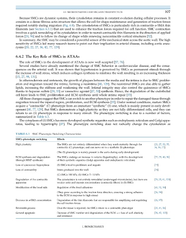
All these changes suggest that SMCs can switch to another phenotype in order to repair the damaged tissue through
migration toward the injured region, proliferation, and ECM synthesis [35]. Under normal conditions, mature SMCs
acquire a “contractile” (C) phenotype from an immature “synthetic” (S) one, which is mainly present in early devel-
opment [40, 77, 129]. But SMCs demonstrate a high plasticity as they are not fully differentiated cells, and they can
return to an (S) phenotype in response to many stimuli. The phenotypic switching is due to a number of factors,
summarized in Table 6.3.
The cytoplasm of (S) SMCs has more developed synthetic organites such as endoplasmic reticulum and Golgi appa-
ratus, leading to hypertrophy [27]. The phenotypic switching does not radically change the cytoskeleton as
TABLE 6.3 SMC Phenotypic Switching Characteristics
SMCs phenotypic switching Effects
High plasticity The SMCs are not entirely differentiated when they reach maturity through the [21, 27, 32, 35,
contractile (C) phenotype, and can move on to a synthetic (S) phenotype 36, 56, 77]
(The (S) phenotype is mainly present in the aorta during early development)
ECM synthesis and degradation The SMCs undergo an increase in volume (hypertrophy), with the development [27, 29, 40, 56]
(through MMP synthesis) of their synthetic organites (Golgi apparatus and endoplasmic reticulum)
Loss of quiescence: hyperplasia (S) SMCs tend to proliferate and migrate [77]
Loss of contractility Stress produced into the wall: [21]
(C) SMCs: 100 kPa; (S) SMCs: 5 10 kPa
Degradation of the contractile The cytoskeleton is not entirely remodeled (undamaged microtubules), but there are [29, 35, 41, 129]
apparatus weaker actin and myosin concentrations (contractile fibers) in (S) SMCs
Modification of the basal side Regulation of the focal adhesions [41, 52, 56]
(They grow according to the traction force direction, ensuring a strong adhesion [24, 96]
to the ECM in response to high stress)
Decrease in α-SMA concentration Degradation of the thin filaments that are responsible for amplifying and regulating [24, 97]
the cell traction forces
Reversible process Once the tissue is repaired, the SMCs return to a contractile phenotype [32, 41, 56]
General apoptosis Decrease of SMC number and degradation of the ECM ¼> loss of wall elasticity [28, 42, 132]
and resistance
I. BIOMECHANICS