
Page 112 - Advances in Biomechanics and Tissue Regeneration
P. 112
108 6. REVIEW OF THE ESSENTIAL ROLES OF SMCS IN ATAA BIOMECHANICS
microtubules remain intact, but the contractile apparatus (i.e., α-SMA thin filaments) is clearly affected [24, 29, 35, 41,
97, 129]. SMC contractility involves a reorganization of their contractile apparatus. In other words, high traction forces
require high adhesion to the ECM; hence, it is suggested that SMCs undergo a regulation of their FAs [24, 52, 96, 133],
evolving toward super focal adhesions (suFAs) in the direction of the applied stress [24, 96].
Hyperplasia concerns the loss of SMC quiescence in favor of a proliferating and migrating behavior [27, 77]. During
hypertension, the increase in wall thickness has been shown to result more from hypertrophy than hyperplasia [31, 98],
but the two phenomena are involved in several pathological states [27]. ATAAs also involve a reduction of the elastin/
collagen ratio in the aortic wall, inducing a stiffness increase and leading to phenotypic switching of SMCs [28]. But the
whole thickness is not uniformly affected: Tremblay et al. [12] have assessed SMC densities across ATAAs and
deduced that it was greater in the outer curvature. The reduced contractile behavior suggests more phenotypic
switching in this area.
6.4.3 SMC Mechanotransduction
As previously highlighted in several studies [21, 68], SMCs tend to remain in a specific mechanical state called
homeostasis. It is considered a reference value for the stress they undergo in the wall under normal physiological
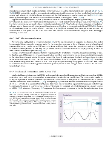
pressure. During any cardiac cycle, SMCs do not activate suddenly their contractile apparatus according to the short
variations of blood pressure. In fact, they always remain partially contracted and tend to adapt gradually to any con-
stant increase of the mean pressure (Fig. 6.9).
Facing a constant rise of wall stress, the SMC response may be divided into two main categories according to time.
In the short term, SMCs react in a progressive contraction until they reach maximal contraction, permitting the regulate
of the blood flow through arterial diameter control. But beyond a given stress threshold, collagen fibers from the
adventitia are recruited to protect the cells and the medial elastic fibers from higher stress values [27, 68]. In the long
term, the remaining mechanical stimuli of SMCs lead to phenotypic switching or apoptosis. In this way, SMCs tend
to coordinate the renewal of ECM, particularly through the synthesis of new collagen fibers to increase the wall resis-
tance to high stress.
6.4.4 Mechanical Homeostasis in the Aortic Wall
Mechanical homeostatis means that SMCs try to regulate their contractile apparatus and their surrounding ECM to
maintain a target wall stress corresponding to a certain mechanobiological equilibrium. The presence of a mechano-
biological equilibrium was first proposed by the constant mean tension of a single MLU in a stressed aorta in spite of
different species and aortic diameter [21, 63]. Humphrey [21] estimated that the circumferential stress per MLU is
about σ θ ¼ 133 kPa. It is assumed that SMCs and fibroblasts tend to maintain a preferred mechanical state through
homeostasis. Kolodney et al. showed that cultured fibroblasts on unloaded gel substrates generate a steady tension
of 3.2 kPa [134]. Moreover, Humphrey [21] suggested that homeostasis expression is similar throughout scales, from
FIG. 6.9 All the smooth muscles do not have the same behavior. The
SMCs are normally partially contracted and adapt their contractile
Cellular force
response and maintain it for a long time. (A) Normally contracted: Sphinc-
ters. (B) Normally partially contracted (tone): blood vessels, airways. A
(C) Phasically active: stomach, intestines. (D) Normally relaxed: esopha-
gus, urinary bladder. Modified from B.M. Koeppen, B.A. Stanton, Berne B
and Levy Physiology, updated edition E-Book, Elsevier Health Sciences, Amster- Contraction SMC
dam, 2009.
C Time
Relaxation D
I. BIOMECHANICS