
Page 360 - Advances in Biomechanics and Tissue Regeneration
P. 360
17.4 BIOMECHANICS AND MECHANOBIOLOGY IN THE CONTEXT OF SKIN WOUND HEALING 357
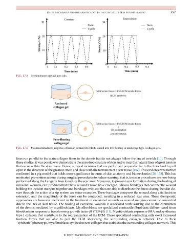
FIG. 17.8 Tension forces applied into cells.
FIG. 17.9 Mechanotransduced response of human dermal fibroblasts loaded into free-floating or anchorage type I collagen gels.
lines run parallel to the main collagen fibers in the dermis but do not always follow the line of wrinkle [10]. Through
these studies, it was possible to demonstrate the anisotropic nature of skin and to map the natural lines of great tension
that occur within the skin tissue. Hence, surgical incisions that are performed perpendicular to the lines tend to pull
open in the direction of the greatest stress and close with the formation of a scar tissue [152]. This evidence was further
confirmed in a pig model that holds more significance in terms of skin anatomy and biomechanics [28, 153]. This has
motivated prevention actions during surgical procedures to reduce scarring, that is, incision procedures are now being
performed along the Langer’s lines to reduce the scar area. Moreover, to prevent scar formation during the healing of
incisional wounds, care products that relieve wound tension have emerged. Silicone bandages that contract the wound
holding the incision margins together and bandages with zip that are able to distribute the forces during the skin clo-
sure through the action of a zip system are some examples. These bandages compress the wound along axial incision
extension, and the magnitude of the force can be controlled, resulting in a reduced scar area. These therapeutic
approaches are however inefficient in the treatment of excisional wounds as wound margins cannot be connected
due to the lack of skin tissue. The healing of excisional wounds is associated with scarring due to the contraction
of the dermis mediated by myofibroblasts. Myofibroblasts are specialized contractile fibroblasts differentiated from
fibroblasts in response to transforming growth factor β1 (TGF-β1) [11]. Myofibroblasts express α-SMA and synthesize
type I collagen that contribute to the reorganization of the ECM. These specialized contracting cells exert increased
traction forces that are able to pull the ECM shortening the surrounding collagen network. Due to their
“synthetic” phenotype, myofibroblasts synthesize new collagen that stabilizes the surrounding collagen network. This
II. MECHANOBIOLOGY AND TISSUE REGENERATION