
Page 193 - Biomedical Engineering and Design Handbook Volume 1, Fundamentals
P. 193
170 BIOMECHANICS OF THE HUMAN BODY
In the tendon excursion method, Euler’s theorem on rotation is used to define an equivalent rela-
tion for the moment arm of a musculotendinous force. Thus, if every change in the relative orienta-
tion of body A and body B can be produced by means of a simple rotation of B in A about the ISA,
then the moment arm of a musculotendinous force can also be written as (Pandy, 1999):
dl M
r M =− A ˆ v B (7.3)
dθ
where l M is the length of the musculotendinous actuator between the effective origin and insertion
sites (P to Q in Fig. 7.14), and q is the angle associated with the simple rotation of body B about the
M
ISA. dl / dθ is the total derivative of musculotendinous length with respect to the angle of rotation
θ , of body B relative to body A.
Equations (7.2) and (7.3) have the same geometric interpretation: the moment arm of a muscle
M
A
x F
force r M , given either by ˆ v B • r OQ ˆ M from Eq. (7.2) or by dl / dθ from Eq. (7.3), is equal to
the perpendicular (shortest) distance between the ISA of B relative to A and the line of action of the
muscle force, multiplied by the sine of the angle between these two lines (Pandy, 1999).
7.5 MUSCLE ACTIVATION AND CONTRACTION DYNAMICS
7.5.1 Muscle Activation Dynamics
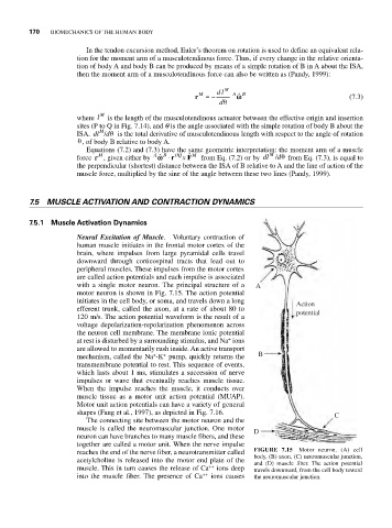
Neural Excitation of Muscle. Voluntary contraction of
human muscle initiates in the frontal motor cortex of the
brain, where impulses from large pyramidal cells travel
downward through corticospinal tracts that lead out to
peripheral muscles. These impulses from the motor cortex
are called action potentials and each impulse is associated
with a single motor neuron. The principal structure of a A
motor neuron is shown in Fig. 7.15. The action potential
initiates in the cell body, or soma, and travels down a long Action
efferent trunk, called the axon, at a rate of about 80 to potential
120 m/s. The action potential waveform is the result of a
voltage depolarization-repolarization phenomenon across
the neuron cell membrane. The membrane ionic potential
+
at rest is disturbed by a surrounding stimulus, and Na ions
are allowed to momentarily rush inside. An active transport
+
+
mechanism, called the Na -K pump, quickly returns the B
transmembrane potential to rest. This sequence of events,
which lasts about 1 ms, stimulates a succession of nerve
impulses or wave that eventually reaches muscle tissue.
When the impulse reaches the muscle, it conducts over
muscle tissue as a motor unit action potential (MUAP).
Motor unit action potentials can have a variety of general
shapes (Fang et al., 1997), as depicted in Fig. 7.16. C
The connecting site between the motor neuron and the
muscle is called the neuromuscular junction. One motor D
neuron can have branches to many muscle fibers, and these
together are called a motor unit. When the nerve impulse
reaches the end of the nerve fiber, a neurotransmitter called FIGURE 7.15 Motor neuron. (A) cell
body, (B) axon, (C) neuromuscular junction,
acetylcholine is released into the motor end plate of the and (D) muscle fiber. The action potential
muscle. This in turn causes the release of Ca ++ ions deep travels downward, from the cell body toward
into the muscle fiber. The presence of Ca ++ ions causes the neuromuscular junction.