
Page 313 - Biomedical Engineering and Design Handbook Volume 2, Applications
P. 313
THE PRINCIPLES OF X-RAY COMPUTED TOMOGRAPHY 291
3. Back-projection of each weighted projection over the three-dimensional reconstruction grid:
2 π D ⎛ Dr Dz ⎞
frs z) = ∫ SO 2 Q β ⎜ ⎜ SO , SO ⎟ dβ
(,
,
D
(
0
SO − s) ⎝ D SO − s D SO − s⎠
The inaccuracies resulting from the approximation associated with this type of reconstruction can be
made diminishingly small with decreasing cone angle, which is typically ~10° for microtomography.
Algorithms that are based upon the Feldkamp principles have found favor in practice because of
their good image quality and fast computational speed. However, the circular path that the source fol-
lows lies in a single plane and consequently projects a limited view of the object. Hence, intuitively
it is observed that the greater the number of planes containing a source point, the more accurate the
reconstruction of the object. To this effect it is possible to state a sufficient condition on the nature of
14
source paths for exact cone-beam reconstruction after the formulation of Smith : “If on every plane
that intersects the object there exists at least one cone-beam source point, then the object can be recon-
structed precisely.” The equivalent statement for a fan-beam reconstruction is “If there exists at least
a fan-beam source on any straight line intersecting an object, an exact reconstruction is possible.”
The scanning schemes that have been adapted to the above principle may involve the spiral/helical
motion that requires lateral displacement with rotation. 15 Extensions of the Feldkamp algorithm, for
16
quite general three-dimensional scanning loci, have been developed by Wang et al. For further discus-
sions on the approximate and accurate cone-beam reconstruction the reader is referred to Ref. 17.
10.4 THE MICROTOMOGRAPHY SYSTEM
10.4.1 The X-Ray Source
The spatial resolution is the principal factor that distinguishes microtomography from conventional
medical tomography. In practice, medical systems must provide very high x-ray fluence in order to
minimize the exposure times. This means that the x-ray source has a relatively large extended emis-
sion area of ~5 mm × 5 mm. The source of x-rays for medical/laboratory systems is created by the
bombardment of a solid metal target (tungsten-rhenium) with a directed high-energy electron beam.
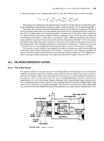
The conventional design produces electrons from a tungsten spiral wire filament (cathode) held at high
voltage. This is heated to a very high temperature, and electrons are extracted in a high voltage field
ΔV formed between the filament housing and the grounded x-ray target (anode) housing (Fig. 10.26).
CA
FIGURE 10.26 Medical x-ray tube.