
Page 314 - Biomedical Engineering and Design Handbook Volume 2, Applications
P. 314
292 DIAGNOSTIC EQUIPMENT DESIGN
The associated geometry acts as an electrostatic lens and focuses the electron beam onto the target.
There is provision for a reduced focal spot size of ~2.5 mm × 2.5 mm from a smaller auxiliary filament
with a corresponding reduction in x-ray emission.
The result of the extended source is a geometric unsharpness in the image that can be interpreted
as that due to a continuous distribution of point sources representing the x-ray emission area. The
extent of the unsharpness U, or blurring, at the edges of the image depends on the extent of the
source, according to
⎛ D ⎞
U = F SD (10.88)
⎜ ⎝ D SO − ⎠ 1 ⎟
where F = source size
D = source to object distance
SO
D = source-to-detector (or image) distance
SD
Hence, if the source size is reduced indefinitely, we have as F → 0 the unsharpness U → 0, and the
magnification M is given by
D
M = SD (10.89)
D SO
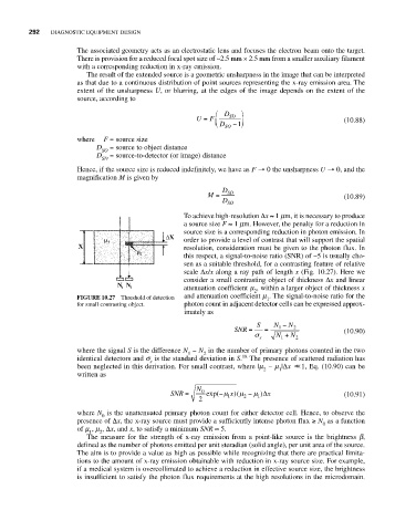
To achieve high-resolution Δx ≈ 1 mm, it is necessary to produce
a source size F ≈ 1 mm. However, the penalty for a reduction in
source size is a corresponding reduction in photon emission. In
order to provide a level of contrast that will support the spatial
resolution, consideration must be given to the photon flux. In
this respect, a signal-to-noise ratio (SNR) of ~5 is usually cho-
sen as a suitable threshold, for a contrasting feature of relative
scale Δx/x along a ray path of length x (Fig. 10.27). Here we
consider a small contrasting object of thickness Δx and linear
attenuation coefficient m , within a larger object of thickness x
2
FIGURE 10.27 Threshold of detection and attenuation coefficient m . The signal-to-noise ratio for the
1
for small contrasting object. photon count in adjacent detector cells can be expressed approx-
imately as
S N − N
SNR = = 1 2 (10.90)
σ s N + N 2
1
where the signal S is the difference N − N in the number of primary photons counted in the two
1 2
identical detectors and s is the standard deviation in S. 18 The presence of scattered radiation has
s
been neglected in this derivation. For small contrast, where |m − m |Δx << 1, Eq. (10.90) can be
2 1
written as
N
SNR = 0 exp( −μ 1 x)(μ 2 − μ 1 )Δ x (10.91)
2
where N is the unattenuated primary photon count for either detector cell. Hence, to observe the
0
presence of Δx, the x-ray source must provide a sufficiently intense photon flux ≥ N as a function
0
of m , m , Δx, and x, to satisfy a minimum SNR ≈ 5.
1 2
The measure for the strength of x-ray emission from a point-like source is the brightness b,
defined as the number of photons emitted per unit steradian (solid angle), per unit area of the source.
The aim is to provide a value as high as possible while recognizing that there are practical limita-
tions to the amount of x-ray emission obtainable with reduction in x-ray source size. For example,
if a medical system is overcollimated to achieve a reduction in effective source size, the brightness
is insufficient to satisfy the photon flux requirements at the high resolutions in the microdomain.