Page 500 -
P. 500
2.5 nm


                                         [001]


                                          [110]         170x170 Å 2

                                      (a)                           (b)
                                              2
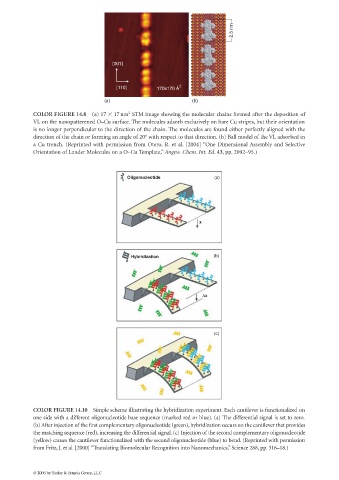
             COLOR FIGURE 14.8 (a) 17   17nm STM image showing the molecular chains formed after the deposition of
             VL on the nanopatterened O–Cu surface. The molecules adsorb exclusively on bare Cu stripes, but their orientation
             is no longer perpendicular to the direction of the chain. The molecules are found either perfectly aligned with the
             direction of the chain or forming an angle of 20° with respect to that direction. (b) Ball model of the VL adsorbed in
             a Cu trench. (Reprinted with permission from Otero, R. et al. [2004] “One Dimensional Assembly and Selective
             Orientation of Lander Molecules on a O–Cu Template,” Angew. Chem. Int. Ed. 43, pp. 2092–95.)


                                              Oligonucleotide               (a)


                                                                       x


                                               Hybridization                (b)


                                                                        ∆x


                                                                            (c)


             COLOR FIGURE 14.10 Simple scheme illustrating the hybridization experiment. Each cantilever is functionalized on
             one side with a different oligonucleotide base sequence (marked red or blue). (a) The differential signal is set to zero.
             (b) After injection of the first complementary oligonucleotide (green),hybridization occurs on the cantilever that provides
             the matching sequence (red), increasing the differential signal. (c) Injection of the second complementary oligonucleotide
             (yellow) causes the cantilever functionalized with the second oligonucleotide (blue) to bend. (Reprinted with permission
             from Fritz, J. et al. [2000] “Translating Biomolecular Recognition into Nanomechanics,” Science 288, pp. 316–18.)


             © 2006 by Taylor & Francis Group, LLC

