
Page 501 -
P. 501
5 µm
COLOR FIGURE 14.11 SEM micrograph of a single E. coli bacterium on an antibody-coated silicon nitride cantilever
oscillator. (Reprinted with permission from Craighead, H.G. [2000] “Nanoelectromechanical Systems,” Science 290,
pp. 1532–35.)
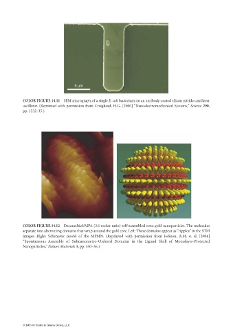
COLOR FIGURE 14.12 Decanethiol/MPA (2:1 molar ratio) self-assembled onto gold nanoparticles. The molecules
separate into alternating domains that wrap around the gold core. Left: These domains appear as “ripples” in the STM
images. Right: Schematic model of the MPMN. (Reprinted with permission from Jackson, A.M. et al. [2004]
“Spontaneous Assembly of Subnanometre–Ordered Domains in the Ligand Shell of Monolayer-Protected
Nanoparticles,” Nature Materials 3, pp. 330–36.)
© 2006 by Taylor & Francis Group, LLC