
Page 330 - Book Hosokawa Nanoparticle Technology Handbook
P. 330
5.5 GRAIN BOUNDARIES AND INTERFACES FUNDAMENTALS
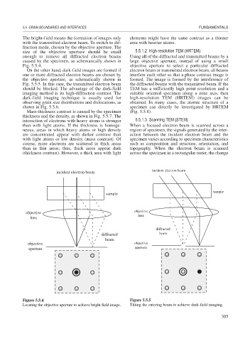
The bright-field means the formation of images only elements might have the same contrast as a thinner
with the transmitted electron beam. To switch to dif- area with heavier atoms.
fraction mode, chosen by the objective aperture. The
size of the objective aperture should be small 5.5.1.2 High-resolution TEM (HRTEM)
enough to remove all diffracted electron beams Using all of the diffracted and transmitted beams by a
caused by the specimen, as schematically shown in large objective aperture, instead of using a small
Fig. 5.5.4. objective aperture to select a particular diffracted
On the other hand, dark-field images are formed if electron beam or transmitted electron beam, all beams
one or more diffracted electron beams are chosen by interfere each other so that a phase contrast image is
the objective aperture, as schematically shown in formed. The image is formed by the interference of
Fig. 5.5.5. In this case, the transmitted electron beam the diffracted beams with the transmitted beam. If the
should be blocked. The advantage of the dark-field TEM has a sufficiently high point resolution and a
imaging method is its high-diffraction contrast. The suitable oriented specimen along a zone axis, then
dark-field imaging technique is usually used for high-resolution TEM (HRTEM) images can be
observing grain size distributions and dislocations, as obtained. In many cases, the atomic structure of a
shown in Fig. 5.5.6. specimen can directly be investigated by HRTEM
Mass-thickness contrast is caused by the specimen (Fig. 5.5.8).
thickness and the density, as shown in Fig. 5.5.7. The
interaction of electrons with heavy atoms is stronger 5.5.1.3 Scanning TEM (STEM)
than with light atoms. If the thickness is homoge- When a focused electron beam is scanned across a
neous, areas in which heavy atoms or high density region of specimen, the signals generated by the inter-
are concentrated appear with darker contrast than action between the incident electron beam and the
with light atoms or low density (mass contrast). Of specimen varies according to specimen characteristics
course, more electrons are scattered in thick areas such as composition and structure, orientation, and
than in thin areas; thus, thick areas appear dark topography. When the electron beam is scanned
(thickness contrast). However, a thick area with light across the specimen in a rectangular raster, the change
incident electron beam incident electron beam
sample
sample
objective objective
lens lens
diffracted
diffracted beam
beam
objective objective
aperture aperture
Figure 5.5.4 Figure 5.5.5
Locating the objective aperture to achieve bright field image. Tilting the entering beam to achieve dark-field imaging.
305