
Page 311 - Vibrational Spectroscopic Imaging for Biomedical Applications
P. 311
Raman Imaging for Biomedical Applications in Clinics 285
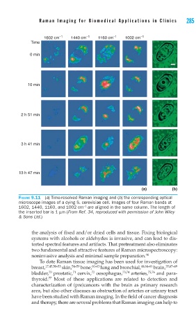
1602 cm –1 1440 cm –1 1160 cm –1 1002 cm –1
Time
0 min
10 min
2 h 51 min
3 h 41 min
13 h 47 min
(a) (b)
FIGURE 9.11 (a) Time-resolved Raman imaging and (b) the corresponding optical
microscope images of a dying S. cerevisiae cell. Images of four Raman bands at
−1
1602, 1440, 1160, and 1002 cm are aligned in the same column. The length of
the inserted bar is 1 μm (From Ref. 34, reproduced with permission of John Wiley
& Sons Ltd.)
the analysis of fixed and/or dried cells and tissue. Fixing biological
systems with alcohols or aldehydes is invasive, and can lead to dis-
torted spectral features and artifacts. That pretreatment also eliminates
two fundamental and attractive features of Raman microspectroscopy:
noninvasive analysis and minimal sample preparation. 46
To date Raman tissue imaging has been used for investigation of
breast, 17,45,50–53 skin, 54–59 bone, 60–63 lung and bronchial, 48,64–66 brain, 35,67–69
72
70
71
bladder, prostatic, cervix, oesophagus, 73,74 arteries, 75,76 and para-
77
thyroid. Most of these applications are related to detection and
characterization of (pre)cancers with the brain as primary research
area, but also other diseases as obstruction of arteries or urinary tract
have been studied with Raman imaging. In the field of cancer diagnosis
and therapy, there are several problems that Raman imaging can help to