
Page 304 - Biomedical Engineering and Design Handbook Volume 2, Applications
P. 304
282 DIAGNOSTIC EQUIPMENT DESIGN
overlap, with the nearest centers of the overlapped region occurring at w =±1/2t, providing 1/2t < W.
Therefore, to avoid overlapping we require
1
τ (10.52)
2W
Hence, by decreasing the sampling interval t, we can separate the transformed functions (Fig. 10.13d).
This means that we can multiply the transform by the function
⎧1 − W w W
Hw() = ⎨ (10.53)
⎩0 elsewhere
−1
to isolate X(w) (Fig. 10.13e). The inverse Fourier transform F {X(w)} of the isolated function will
yield the original continuous function x(t) (Fig. 10.13f). The complete recovery of a band-limited
function, from samples whose spacing satisfies Eq. (10.51), has been formulated as the Whittaker-
Shannon sampling theorem. If the condition of Eq. (10.51) is not met, the transform in the interval
(−W, W) is corrupted by contributions from adjacent periods. This leads to a phenomenon known as
aliasing and prevents the complete recovery of an undersampled function. 12
Shannon’s sampling theorem is closely related to the Nyquist criterion. With respect to telegraph
transmission theory, Nyquist proposed the basic idea that the minimum signal rate must contain the
bandwidth of the message. For sampled signals, the bandwidth really ranges from −f /2 to f /2, where
s s
f is the sampling rate. So the minimum sampling rate f (known as the Nyquist rate), does contain
s s
the signal bandwidth, whose highest frequency is limited to the Nyquist frequency f . Shannon gives
s
credit to Nyquist for pointing out the fundamental importance of the time interval 1/2W in telegra-
phy. However, he further established that if a function f(t) contains no frequencies higher than W, it
is completely determined by samples at a series of points spaced W/2 apart.
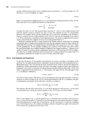
10.3.4 Line Integrals and Projections
To describe the process of tomographic reconstruction, we need to formulate a description of the
geometry of x-ray paths and the process of absorption along the paths that result in the projected two-
dimensional image. This will provide the basic framework from which to develop the method for
building up a digital representation of the solid object. We define a single ray path as the straight line
joining the x-ray source to a detector cell and passing through the absorbing object (Fig. 10.14). The
object is represented by the function f(x, y). The location r and angle q of ray AB with respect to the
(x, y) coordinates is defined by
x cosθ + ysinθ = r (10.54)
The level of input signal to the detector cell is determined by the integrated absorption of photons
along the ray path according to Eq. (10.22). For the path defined by the (q, r) coordinates, the pho-
ton attenuation, over length l of ray AB in the s direction, is written as
( ∫
Φ () = Φ exp − f ( , )xyds ) (10.55)
r
θ
0
AB
This indicates that the object function f(x, y) is the linear attenuation coefficient m(x, y) in the plane
of the projected rays. Taking the natural logarithm of Eq. (10.55) gives the linear relation
Φ r ()
Pr() =− ln θ = ∫ f xyds (10.56)
(,
)
θ
Φ 0 AB
This is a line integral of f(x, y) and the quantity P (r) is called the radon transform or the projection
q
of f(x, y) along the ray path. It is important to note that the process of reconstructing slices of objects
from their projections is simply that of inverting Eq. (10.56). A projection is formed by combining
a set of line integrals P (r), the simplest one being a collection of parallel ray paths with constant q.
q
This is known as a parallel projection (Fig. 10.15).